
CULTURE SPHEROIDS
3D cell can be cultured directly in a ULA, round bottom plate to develop the typical spherical shape of a spheroid. Cells can also be grown in alternate labware like a petri dish, flask, or other microplate before transferring to an imaging microplate.
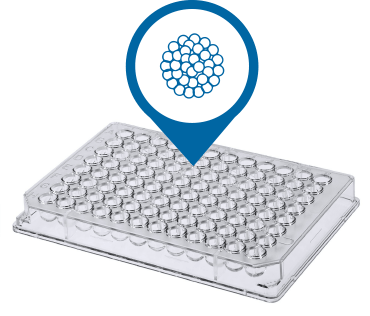
96- or 384- Well Plates
Most 3D cell cultures for high-content, high-throughput screening are run using 96- or 384-well microplates. ULA, U-shaped/round bottom microplates enable the spherical shape to form spheroids or spheroids can also be cultured in a flat-bottom microplate using hydrogels to simulate extracellular matrices (ECM). Optimized spheroid cell culture protocols for the ImageXpress® Micro Confocal High-Content Imaging System can use ULA, U-bottom 96- and 384-well plates with a one-step staining procedure that reduces assay time and minimizes variability.
Shopping for reagents?
Check out our
EarlyTox Cell Viability Assay Kits

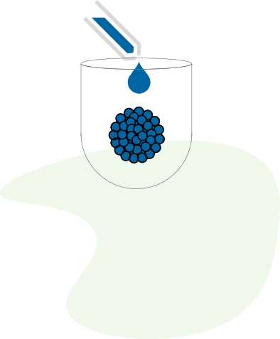
TREAT WITH COMPOUNDS
After spheroid formation, compounds are added into the wells, and then incubated for one to several days, depending on the mechanism being studied. Generally, shorter durations are used to study apoptosis and longer durations for multi-parameter cytotoxicity studies. For drug treatments requiring a longer duration, compounds are refreshed periodically during incubation. The concentration of compounds and the incubation period is dependent on the desired protocol.

STAIN FOR MARKERS
After compound treatment is completed, stains are added directly to the media. Stains that require no washing can be used to avoid disturbing spheroids, but spheroids can be carefully washed even using automation, if necessary.

Assay Kits
Easy-to-use, robust assay kits for life science research, drug discovery and development, and bioassays. Our assay kits are optimized for use on our instruments. Screen more compounds earlier in drug discovery and enable characterization of a full concentration-response profile of test compounds for cell viability, cell proliferation, axiogensis, and more.
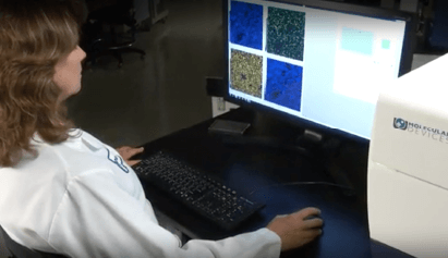
High-content Imaging System
The ImageXpress Micro Confocal system is a unique confocal imaging solution capable of imaging more than a million wells a week. MetaXpress software enables acquisition of 3D z-stacks. Z-plane images can be saved individually or collapsed into a single 2D projection image using a mathematical algorithm.

ACQUIRE 3D CELL IMAGE
Images within the body of the spheroid can be captured individually or as a z-stack (multiple images taken at differing depths) using specialized imaging equipment.


ANALYZE 3D CELL IMAGE
Use cellular imaging analysis software to run quantitative analysis of the cell images to assess and monitor how cells express against different markers and to quantify biological readouts.
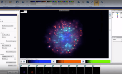
High-content Imaging Software
MetaXpress High-Content Image Acquisition and Analysis Software is a multi-level analysis tool for a wide range of 2D and 3D applications optimized for the ImageXpress Micro Confocal system. The integrated acquisition and analysis application modules for 3D cell models simplify high-throughput quantification of 3D structures with volume, intensity, and distance measurements.