Organoids
Biologic research and disease modeling recapitulate complexity of real tissues
What are organoids?
Organoids are three-dimensional (3D) multi-cellular, microtissues derived from stem cells that are designed to closely mimic the complex structure and functionality of human organs like the lung, liver or brain. Organoids are multi-cellular and demonstrate a high order of self-assembly to allow for an even better representation of complex in vivo cell responses and interactions, as compared to traditional 2D cell cultures.
There are three distinct definitions that differentiate an organoid:
- It's a 3D biological microtissue containing several types of cells
- Represents the complexity, organization and structure of a tissue
- And, resembles at least some aspect of a tissue’s functionality
Organoids are becoming increasingly important in the fields of cancer research, neurobiology, stem cell research, and drug discovery, since they allow for the enhanced modeling of human tissues. Derived from stem cells, organoids can be differentiated into a wide range of tissue types including liver, lung, brain, kidney, stomach, and intestine. Because these 3D microtissues mimic in vivo organs, they can provide researchers with greater insight into the mechanisms of human development and disease. For example, researchers can grow organoids from genetically modified cells to understand how specific gene mutations are linked to certain genetic disorders. Organoids can also facilitate the study of infectious diseases and host-pathogen interactions. Finally, the ability to use patient-derived organoids for drug screens and toxicity evaluations is enabling researchers to make further advancements in personalized medicine.
General workflow for organoid culturing and screening
Due to the growing complexity of organoids and other 3D cellular systems, more sophisticated 3D imaging and analysis techniques are needed to accurately and efficiently characterize these biological assays. Today, automated confocal imaging systems and 3D image analysis software are commonly used to help researchers streamline their workflow and obtain optimal results.
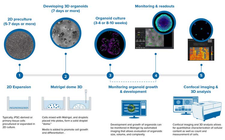
Step 1) 2D preculture
Organoids are derived from either primary cells (i.e., intestine, lung, or kidney) or induced pluripotent stem cells. Stem cells are able to differentiate and self-assemble into a variety of tissue-specific organoids.
Step 2) Developing 3D organoids
Typically cells are premixed with Matrigel and droplets are placed into a 24-well plate at room temperature. The plates are then placed into an incubator to form a solid droplet dome. Media is then added for seven or more days to promote cell growth and differentiation into specific tissue (brain, gut, lungs, etc.). Media includes ECM proteins, and different growth factors, which will vary depending on the type of tissue that is being developed.
Step 3) Organoid culture
Organoid culture is a long process and may include several steps with different media. During this process, cell health needs to be monitored (imaging), typically used for understanding developmental biology and tissues.
Step 4) Monitoring and readouts
Before experiments are conducted, organoids need to be monitored and characterized to ensure that they have the appropriate tissue structure and differentiation. High-content imaging allows for monitoring and visualizing the growth and differentiation of organoids, 3D reconstruction of the structures, complex analysis of organoids structure, cell morphology and viability, as well as the expression of different cell markers.
Step 5) Confocal imaging and 3D analysis
Confocal imaging and 3D analysis of organoids allow visualization and quantitation of the organoids and the cells that make up the organoid. Characterization of multiple quantitative descriptors of organoids could be used for studying disease phenotypes and compound effects.
Confocal imaging and 3D image analysis of organoids
Organoids are very useful for disease modeling and assessment of compound effects. Automated imaging and analysis of organoids are important for the quantitative assessment of phenotypic changes in organoids, as well as for increasing throughput of experiments and screens.
Confocal imaging, like the ImageXpress® Confocal HT.ai system with high-performance lasers and water immersion objectives are especially useful for capturing the complexity of 3D biological assays. Unlike typical spheroids, which have appearance of solid objects and limited penetration of light, some 3D organoids such as pulmonary organoids have a hollow appearance, with lumen, or cavities inside, and are more easily penetrated by light, allowing “imaging through” the microtissues embedded into Matrigel.
High-content analysis tools like the MetaXpress or IN Carta Image Analysis Software, tools allow for the finding and characterization of multiple objects/organoids, either in 2D format (for single plane, or maximum projection images) or in 3D when objects from the multiple planes are connected and reconstituted in 3D space by software. Organoids can be characterized for diameter, volume, shape, intensity of specific marker, or distance to other objects.
Furthermore, individual cells, nuclei, or organelles can be defined and measured within each organoid. That allows count of live and dead cells, or cells with a specific marker also defining volumes and distances between the objects. Numeric values can be counted for each organoid or averaged per well.
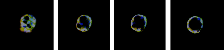
Lung organoid cell image gallery
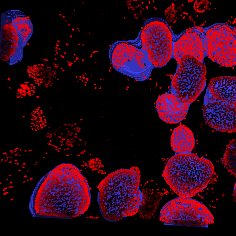
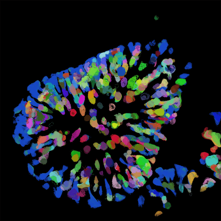
Image analysis of organoid
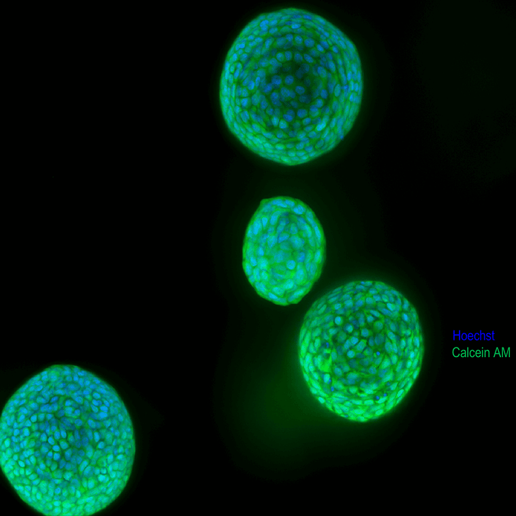
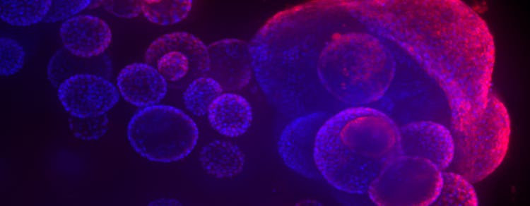
Lung organoids
Lung organoids 2

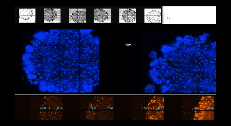
Automated imaging of live organoids in Matrigel was done using confocal option, 4x or 10x magnification
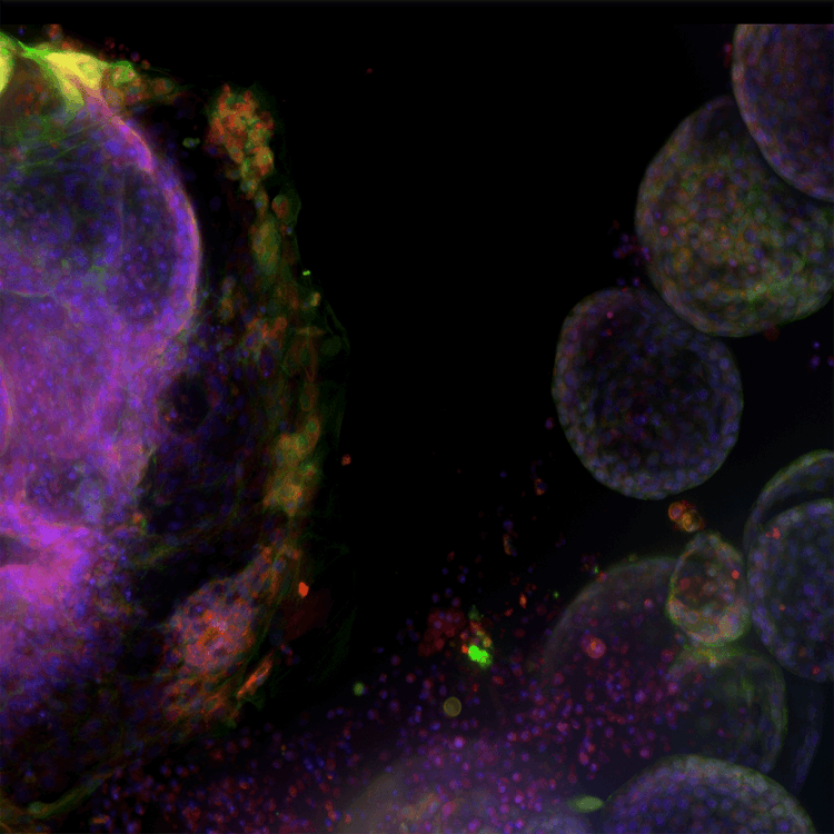
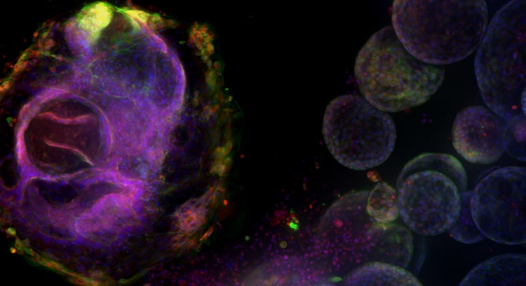
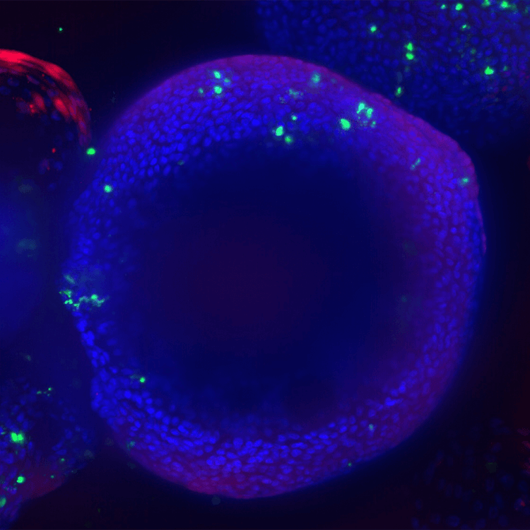
High complexity lung organoid
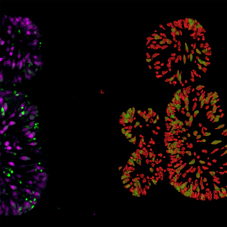
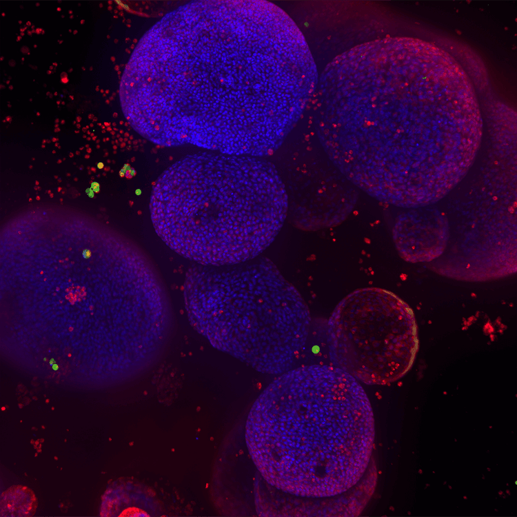
Gut organoid dTomato cells in red Detection of Double positive(mNeon) in green
Internally expressed genes dTomato magenta mNeon green 10x
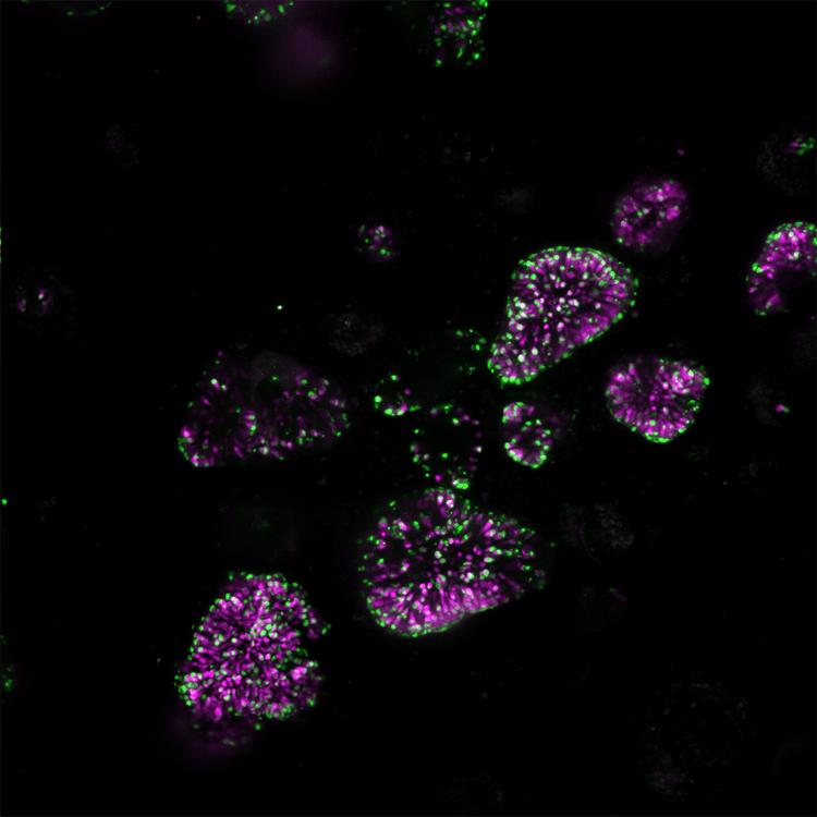
Intestinal organoid 40x,dTomato cell count
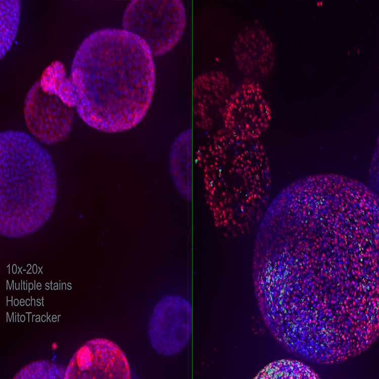
Lung organoid 10x 20x Multiple stains Hoechst MitoTracker
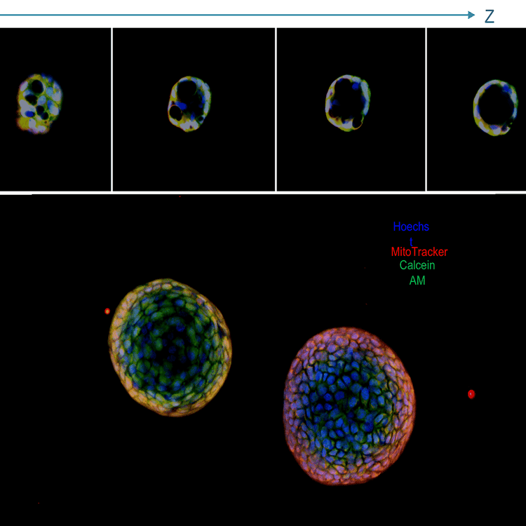
Lung organoids,projection image and a panel with Z stack; live stained with Hoechstblue,nuclei),MitoTracker(red,mitochondria),and Calcein AM(green,viability dye);20x water immersion
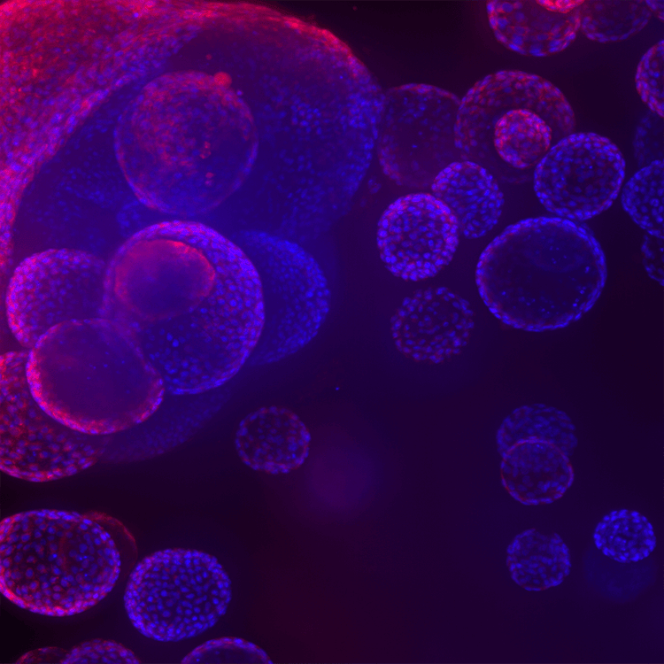
Lung organoids 3
Lung organoids 4
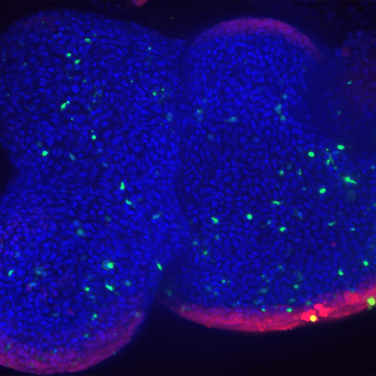
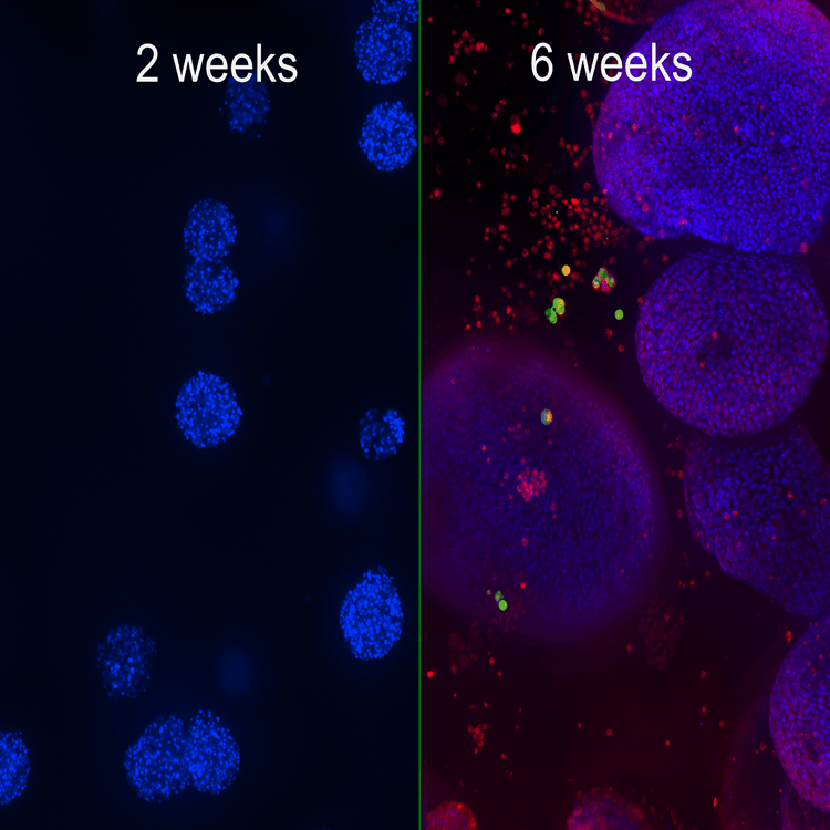
Lung organoids lung airway epithelial cells cultured in matrigel domes for 8 weeks
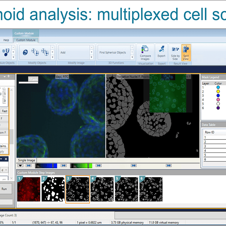
Organoid analysis multiplexed cell scoring
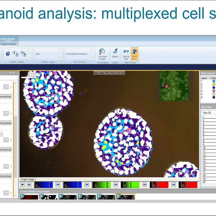
Organoid analysis multiplexed cell scoring 2
Organoid development